Understanding Your Luteal Phase Defect, and What It Means for Conception
The 2am Google Search You Know Too Well
You have been tracking your cycle for months. You chart your basal body temperature before you even open your eyes each morning. You use LH strips every cycle. You know your ovulation day better than your own birthday.
And yet, something is persistently, quietly wrong.
Your period keeps arriving too early. Or there is that frustrating spotting, brownish and unwelcome, appearing three, four, sometimes five days before your actual bleed. Your luteal phase temperature rise drops too soon. And when you finally typed “short luteal phase” into a search engine at midnight, your stomach sank a little, because everything you read seemed to confirm what you had suspected for a long time.
You visited your GP. They ran day-21 progesterone and told you it was “normal.” They suggested you relax. They said you were young enough not to worry.
But you are still not pregnant. Or you have been pregnant, briefly, and you have lost it. And you are starting to wonder whether the answer everyone keeps dismissing is actually the answer.
It may well be. What you are experiencing could be a luteal phase defect. It is one of the most frequently missed contributors to difficulty conceiving and to recurrent early pregnancy loss. It is also, crucially, one of the most treatable hormonal conditions in reproductive medicine, once it has been properly identified and addressed.
You are not imagining this. Your instincts deserve to be taken seriously.
What Is a Luteal Phase Defect? The Clinical Foundation
Featured snippet answer: A luteal phase defect is a condition in which the second half of the menstrual cycle, the luteal phase, is either too short (fewer than 10 days) or characterised by insufficient progesterone production. Both scenarios prevent the uterine lining from developing adequately, impairing embryo implantation and increasing the risk of very early pregnancy loss. It is one of the most underrecognised hormonal causes of unexplained infertility and recurrent miscarriage.
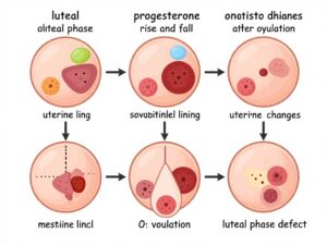
The luteal phase begins the day after you ovulate and ends the day your period starts. During this phase, a temporary structure called the corpus luteum forms in your ovary from the collapsed follicle that released your egg. The corpus luteum has one primary job: to produce progesterone.
Progesterone is the hormone that transforms the uterine lining from a freshly built structure into a fully furnished home, ready to receive a fertilised embryo. Think of oestrogen, which dominates the first half of your cycle, as the architect and builder. Progesterone is the interior designer who arrives after the walls are up, adds warmth, blood supply, and receptivity, and makes the space genuinely liveable for a new life.
A luteal phase defect (also referred to as luteal phase insufficiency or luteal phase deficiency) occurs when one of two things goes wrong. Either the luteal phase is too short for implantation to succeed, typically under 10 days, or progesterone production during the luteal phase is inadequate, even when the timing appears normal on a chart.
What makes this condition so difficult to detect is that there is no single gold-standard diagnostic test, and progesterone testing is frequently performed at the wrong point in the cycle. This is not a rare edge case. It is a systemic gap in the way reproductive hormonal health is assessed in standard primary care.
The Luteal Phase Day by Day: What Healthy Hormones Are Actually Doing
To truly understand a luteal phase defect, you need to understand what a healthy luteal phase looks like, because the detail matters far more than most fertility tracking apps acknowledge.
After ovulation, the follicle that held your egg collapses inward and transforms into the corpus luteum. This small, intensely active gland begins producing progesterone almost immediately. Progesterone levels rise steeply over the first several days after ovulation, reaching their peak around days five to seven of the luteal phase.
Simultaneously, the uterine lining, already thickened by oestrogen in the first half of your cycle, undergoes a critical transformation. Under the influence of progesterone, the endometrial cells differentiate and become receptive to a fertilised embryo. The glands within the lining begin secreting nutritional fluid that the early embryo will need before it establishes a blood supply. This extraordinary, tightly timed process is called the implantation window, and it typically occurs between days six and ten after ovulation.
If fertilisation does not occur, the corpus luteum gradually deteriorates. Progesterone falls. The uterine lining sheds. Your period begins. This natural decline is supposed to happen. The problem arises when it happens too early.
If fertilisation does occur, the embryo begins producing human chorionic gonadotrophin, or hCG, the hormone detected by pregnancy tests. This hCG signals the corpus luteum to continue producing progesterone rather than shutting down, effectively rescuing it from its scheduled demise. The corpus luteum then supports progesterone production for approximately the first 8 to 10 weeks of pregnancy, until the placenta is mature enough to take over this function entirely.
This is why the luteal phase is so consequential for conception. The window between ovulation and your expected period must be long enough, and hormonally rich enough, to allow implantation to succeed and those first fragile days of a pregnancy to be supported. When either condition fails, even a successfully fertilised embryo may be lost before you ever receive a positive test.
How Common Is a Luteal Phase Defect?
This is a question that deserves a straight answer, and the honest response is that we do not have a precise figure, and that uncertainty is itself revealing.
Research suggests that luteal phase defect may be present in approximately 3 to 10 percent of women experiencing infertility. In women with recurrent pregnancy loss, rates as high as 35 percent have been reported, though these figures likely underestimate the true prevalence because of inconsistent diagnostic criteria and the absence of routine mid-luteal hormone assessment.
Certain groups are at increased risk. Women in their late 30s and early 40s experience declining corpus luteum function as part of natural reproductive ageing. Women with polycystic ovary syndrome, PCOS, often have irregular ovulation that produces a suboptimal corpus luteum. Women with hyperprolactinaemia, meaning elevated levels of the hormone prolactin, have disrupted LH signalling that impairs corpus luteum function downstream. Thyroid disorders, particularly hypothyroidism, are also closely associated with luteal phase insufficiency.
Women who exercise intensely, restrict calories significantly, or carry a significant chronic psychological stress load are also at elevated risk, because all of these factors interfere with the hypothalamic-pituitary-ovarian axis, which is the central command chain that governs your entire hormonal cycle.
Finally, women who have undergone fertility treatments involving ovarian stimulation drugs are at particular risk of luteal phase deficiency in treatment cycles. The medications used to trigger ovulation suppress the natural corpus luteum’s ability to function, which is precisely why progesterone supplementation is standard practice in IVF and other assisted reproduction protocols.
Knowing where you sit within this picture helps you evaluate your own situation with clear eyes, rather than dismissing symptoms because a single routine test appeared unremarkable.
Recognising a Luteal Phase Defect: The Symptoms That Deserve Attention
Many online resources describe only the most obvious presentations of luteal phase defect: a short cycle, spotting before a period, difficulty getting pregnant. But the clinical picture is frequently more nuanced, and several presentations are consistently missed.
A luteal phase consistently under 10 to 12 days. If you have been tracking your cycles and repeatedly observe that ovulation occurs but your period arrives fewer than 10 days later, this is the most direct indicator of a shortened luteal phase. Anything under 10 days is considered clinically significant. Even a luteal phase of 10 or 11 days can be borderline if progesterone levels are not adequately elevated during it.
Pre-menstrual spotting beginning more than 2 days before your period. A day or two of light spotting before a period is common and generally unremarkable. But persistent brown or pink spotting beginning 3, 4, or 5 days before your bleed is worth investigating. This often reflects premature progesterone withdrawal, causing the uterine lining to begin shedding before it should.
Recurrent chemical pregnancies or very early losses. A chemical pregnancy is a positive pregnancy test followed by a bleed before a heartbeat is ever detected on ultrasound, typically occurring within the first few days of a missed period. These losses are heartbreaking in part because they are so frequently unacknowledged. Many women experience multiple chemical pregnancies without ever receiving an explanation. A luteal phase defect is a significant and plausible contributor, because inadequate progesterone support in the implantation window can allow an embryo to implant but fail to sustain.
Overall short menstrual cycles. A cycle length consistently below 24 to 25 days, particularly when the follicular phase is of normal length, often reflects a shortened luteal phase rather than early ovulation. If your overall cycles are brief, it is worth calculating what proportion of your cycle is luteal phase and whether that proportion is adequate.
Disproportionately intense premenstrual symptoms. There is growing evidence that some women with luteal phase defect experience pronounced PMS, including breast tenderness, mood disruption, irritability, and bloating, not because of elevated progesterone but because progesterone fluctuates erratically rather than maintaining a steady, supported peak before its natural decline. This can be dismissed for years as simply “bad PMS” when it actually reflects hormonal dysregulation worth investigating.
A history of early pregnancy losses at or around the expected period. Some women conceive regularly but miscarry in the first four to six weeks, sometimes before a clinical pregnancy has even been confirmed on ultrasound. When this pattern recurs across multiple cycles, it constitutes a consistent clinical signal. A luteal phase defect is one of the most treatable explanations for this pattern, and yet it is among the last things investigated.
Difficulty maintaining pregnancies despite confirmed conception. If you have found it harder to progress beyond a positive test to a confirmed heartbeat, regardless of how long you have been trying, the quality of your luteal phase is a reasonable and clinically important question to pursue.
The Root Causes of Luteal Phase Defect: Why This Happens to You
Luteal phase defect is rarely a standalone problem. It is almost always a consequence of something else disrupting the hormonal signalling chain. Understanding the underlying cause is what makes treatment both possible and sustainable. Here is what the evidence currently supports.
Suboptimal follicular development in the first half of your cycle. The corpus luteum is only as good as the follicle that produced it. If the follicle did not develop adequately before ovulation, due to insufficient FSH stimulation, poor follicular response, or conditions like diminished ovarian reserve, the resulting corpus luteum will be similarly compromised. This is one of the most common and least-discussed mechanisms behind luteal phase defect, and it is why addressing the follicular phase, not just the luteal phase, is often central to treatment.
Elevated prolactin. Prolactin is the hormone primarily associated with breast milk production, but elevated prolactin outside of breastfeeding, a condition called hyperprolactinaemia, disrupts reproductive hormone signalling in multiple ways. High prolactin suppresses GnRH (gonadotrophin-releasing hormone) from the hypothalamus, which reduces FSH and LH from the pituitary, which in turn compromises both ovulation quality and corpus luteum function. Hyperprolactinaemia can stem from a small benign pituitary tumour called a prolactinoma, from thyroid disease, from certain medications, or from chronic psychological stress.
Thyroid dysfunction. The thyroid gland and the reproductive hormonal axis are intimately connected. Even subclinical hypothyroidism, where TSH is elevated but still within the conventional “normal” reference range, can impair progesterone production, affect the liver’s clearance of oestrogen, and reduce endometrial receptivity. Many women with luteal phase defect find that addressing an underlying thyroid imbalance resolves or substantially improves their luteal function, without any direct progesterone supplementation required.
Chronic stress and the cortisol-progesterone competition. The hypothalamic-pituitary-adrenal, or HPA, axis governs your body’s stress response. Under chronic stress, cortisol levels remain persistently elevated. Both cortisol and progesterone are steroid hormones synthesised from a shared precursor molecule called pregnenolone. When the body is under sustained stress, it prioritises cortisol production, effectively diverting the raw material that would otherwise go towards progesterone synthesis. This mechanism, sometimes called the “pregnenolone steal,” is one of the most underappreciated pathways in reproductive endocrinology and one of the most actionable once it is understood.
High-intensity exercise combined with insufficient caloric intake. Women who exercise intensely while eating in a significant caloric deficit can develop relative energy deficiency, which disrupts the pulsatile GnRH release from the hypothalamus that drives the entire reproductive hormone cascade. The result is suppressed LH, impaired ovulation quality, and a weakened corpus luteum. This pattern is not exclusive to professional athletes. It can occur in any woman whose energy availability is chronically below what her body requires to maintain reproductive function alongside everything else.
Age-related changes in ovarian function. As women move through their mid-to-late 30s, follicle quality and quantity begin to decline, and ovulation becomes less consistently robust. Corpus luteum function may diminish correspondingly. This is not a reason for despair. It is a physiological reality that can be meaningfully supported with targeted interventions, but it does require earlier and more proactive investigation than younger women typically receive.
The use of NSAIDs around ovulation. Non-steroidal anti-inflammatory drugs, including ibuprofen, can inhibit the prostaglandins required for follicle rupture and corpus luteum formation when taken in the days surrounding ovulation. This is a rarely discussed but clinically important contributor, particularly for women who routinely take ibuprofen for pain management.
Diagnosing a Luteal Phase Defect: The Tests Worth Requesting From Your Doctor
One of the most common sources of frustration for women with luteal phase defect is receiving apparently normal test results while their symptoms persist. The problem is frequently not the results themselves, but when and how the tests are ordered.
Serum progesterone 7 days post-confirmed ovulation. The standard day-21 progesterone test is only accurate if your cycle is 28 days long and you ovulate on day 14. If your cycle is longer, shorter, or irregular, a day-21 test may capture you at entirely the wrong point in your luteal phase. A far more accurate approach is to draw progesterone 7 days after confirmed ovulation, using LH strip testing or basal body temperature charting to establish when ovulation actually occurred. A mid-luteal progesterone above 30 nmol/L is generally considered adequate, though some reproductive specialists prefer levels above 35 to 40 nmol/L for optimal implantation support.
Serial progesterone measurements. Progesterone is secreted in pulses, meaning a single reading can be misleading in either direction. Drawing progesterone on two or three occasions within the mid-luteal phase and examining the overall pattern, rather than any single value, provides a much more complete and clinically meaningful picture.
Luteal phase length documentation. Tracking your cycle across three to six months using LH strips and BBT charting, and consistently observing a luteal phase below 10 days, is itself diagnostically valuable. Bringing detailed charts to a clinical appointment gives a specialist more useful information than almost any single blood draw.
Full thyroid panel. A TSH alone is insufficient when investigating luteal phase defect. Request free T3, free T4, and thyroid antibodies (anti-TPO and anti-Tg), because thyroid autoimmunity can affect reproductive outcomes even when TSH appears normal.
Fasting prolactin. This is a blood test best drawn in the morning, after a night of adequate sleep and without recent breast stimulation or vigorous exercise, as these can all artifically elevate the result. Elevated prolactin is a treatable cause of luteal phase defect and is straightforward to screen for.
Day-2 or day-3 FSH, LH, and oestradiol. These early-cycle hormone levels provide information about ovarian reserve and the quality of follicular stimulation your ovaries receive each cycle. They help contextualise whether a suboptimal corpus luteum is a downstream consequence of inadequate follicular development.
You have every right to request this workup. These are not unusual or specialist-only tests. They form the minimum reasonable investigation for a woman presenting with cycle abnormalities and difficulty conceiving, and you should feel empowered to ask for them by name.
8 Evidence-Based Strategies for Supporting Conception With a Luteal Phase Defect
Once a luteal phase defect has been identified, there are genuinely effective, evidence-supported options available. The most appropriate approach will depend on your specific underlying cause, and specialist involvement is important for personalising your plan. But here is what the current evidence supports.
1. Progesterone Supplementation After Confirmed Ovulation
Mechanism: Supplementary progesterone, taken after confirmed ovulation, directly compensates for what the corpus luteum is not producing adequately. It can be administered vaginally, which is the most commonly used and best-tolerated route, or orally. The goal is to support endometrial development and maintain the uterine lining through the implantation window and into the early weeks of pregnancy.
Evidence level: Clinical consensus holds that vaginal progesterone supplementation is effective for luteal phase support, and it is standard practice in assisted reproduction worldwide. For natural conception cycles with confirmed luteal phase defect, the evidence base is smaller but supportive. Research suggests that vaginal micronised progesterone initiated after confirmed ovulation, and continued until 10 to 12 weeks of a confirmed pregnancy, significantly reduces early pregnancy loss in women with a documented history of recurrent miscarriage.
Practical note: This is a prescription treatment. Timing, dose, and duration are clinically important variables. Beginning supplementation before ovulation has been confirmed may suppress ovulation in some cycles, which defeats the purpose entirely. Work with a gynaecologist or reproductive endocrinologist to establish the correct protocol for your situation.
2. Treating Hyperprolactinaemia at Its Source
Mechanism: When elevated prolactin is confirmed as the underlying driver of luteal phase defect, normalising prolactin levels restores the GnRH-FSH-LH signalling cascade. This allows adequate follicular development and robust corpus luteum formation to resume, improving progesterone production naturally without requiring direct progesterone supplementation.
Evidence level: Clinical consensus holds that treating hyperprolactinaemia with dopamine agonist medications restores ovulatory function and luteal adequacy in the majority of affected women. Conception rates improve meaningfully once prolactin is within the normal range.
Practical note: Hyperprolactinaemia requires proper investigation of its cause before treatment. An MRI of the pituitary may be recommended to exclude a prolactinoma. This is managed collaboratively by an endocrinologist and a reproductive specialist.
3. Thyroid Optimisation Before and During Conception
Mechanism: Ensuring thyroid function is within the optimal range for reproduction, rather than merely within the broad reference range used in general medicine, supports progesterone synthesis, endometrial receptivity, and immune tolerance of early pregnancy. Research suggests that a TSH below 2.5 mIU/L is associated with better fertility and lower miscarriage rates.
Evidence level: There is growing evidence that subclinical hypothyroidism, particularly when accompanied by elevated thyroid antibodies (as in Hashimoto’s thyroiditis), contributes to implantation failure and early pregnancy loss. Several professional bodies, including the American College of Obstetricians and Gynecologists, recommend thyroid optimisation before and during conception in women with thyroid abnormalities.
Practical note: If your TSH is between 2.5 and 4.0 mIU/L and you have thyroid antibodies, discuss whether a trial of low-dose levothyroxine before conception is appropriate with your GP or reproductive specialist. This conversation is worth initiating proactively.
4. Restructuring the Stress Burden at a Physiological Level
Mechanism: Reducing chronic activation of the HPA axis frees up pregnenolone for progesterone synthesis rather than cortisol. This is not a suggestion to “stress less” as if that were simply a decision. It is about making concrete structural changes to how your nervous system is operating day to day, particularly during your luteal phase.
Evidence level: Research suggests that mind-body interventions including mindfulness-based stress reduction, yoga, and cognitive behavioural therapy can reduce cortisol and improve reproductive outcomes in women with infertility. The evidence base is growing, though studies are still limited by small sample sizes and methodological variability.
Practical note: The most meaningful changes are consistent rather than sporadic. Prioritising 7 to 9 hours of sleep nightly, moderating high-intensity exercise during the luteal phase, and addressing sources of sustained psychological pressure are all interventions with measurable hormonal effects. These are concrete clinical recommendations, not vague wellness advice.
5. Supporting Follicular Development in the First Half of Your Cycle
Mechanism: Because the quality of the corpus luteum depends directly on the quality of the follicle that preceded it, improving follicular development in the first half of your cycle, the follicular phase, directly improves luteal function. Ovulation induction with medications such as letrozole or clomiphene citrate can improve follicular development in women with ovulatory dysfunction, and in doing so, produce a more competent corpus luteum.
Evidence level: Clinical consensus holds that ovulation induction improves luteal function in women with ovulatory irregularities. There is also research supporting the use of low-dose FSH injections in women with subtle ovulatory deficiencies who have not responded adequately to oral agents.
Practical note: Ovulation induction requires monitoring by a specialist and carries a risk of multiple pregnancies. It is most appropriate for women with ovulatory dysfunction as a contributing factor, rather than for women who ovulate normally but have isolated luteal phase insufficiency.
6. Targeted Nutritional Support for Hormone Synthesis
Mechanism: Several micronutrients play documented roles in steroid hormone synthesis and endometrial health. Vitamin D acts as a precursor within the steroidogenesis pathway and has receptors in both ovarian and endometrial tissue. B vitamins, particularly B6, support progesterone receptor sensitivity and the conversion of progesterone from its precursor molecules. Magnesium is required for over 300 enzymatic processes, including several within the steroid hormone synthesis pathway. Zinc supports LH signalling and progesterone production at the corpus luteum.
Evidence level: Research suggests that vitamin D deficiency is associated with lower progesterone levels and poorer fertility outcomes. There is growing evidence linking B6 and magnesium insufficiency with impaired luteal function, though the independent evidence base for these remains emerging.
Practical note: Testing your vitamin D level is a reasonable and inexpensive starting point. Many women in the UK and across northern latitudes, as well as women with darker skin tones, have suboptimal levels without knowing it. Supplementing to reach an optimal range of 75 to 150 nmol/L is low-risk and may offer meaningful benefit.
7. Reviewing Medications That May Be Impairing Luteal Function
Mechanism: Certain medications can interfere with corpus luteum formation or function. NSAIDs taken in the days surrounding ovulation inhibit prostaglandin synthesis, which is required for follicle rupture and successful corpus luteum formation. Medications that elevate prolactin, including some antipsychotics, certain antidepressants, and gastroprotective drugs such as metoclopramide, can suppress LH and impair progesterone production indirectly.
Evidence level: Clinical consensus holds that NSAIDs should be avoided around ovulation in women trying to conceive. The association between specific medications and elevated prolactin is well-established.
Practical note: Do not discontinue any prescribed medication without speaking to your doctor. But if you are taking regular NSAIDs or any of the above-mentioned drug classes, raising this in the context of your fertility investigation is entirely appropriate.
8. Optimising How You Track Ovulation and Measure Luteal Length
Mechanism: It is impossible to meaningfully assess your luteal phase without first knowing precisely when ovulation occurred. Cycle-length estimates alone are unreliable. LH testing strips used daily from cycle day 10 onward, combined with BBT charting to confirm the post-ovulatory temperature rise retrospectively, give a much more accurate picture of your cycle’s internal architecture.
Evidence level: Research supports urinary LH testing as a reliable predictor of ovulation. BBT charting complements LH testing by providing independent retrospective confirmation and helping to identify patterns across multiple cycles.
Practical note: Record three to six full cycles, noting the day of your LH surge, the day of your post-ovulatory temperature rise, and the first day of your next period. The number of days from confirmed ovulation to your period is your luteal phase length. A persistent luteal phase under 10 days, confirmed across multiple cycles, is clinically meaningful and warrants formal investigation.
The Connection Between Luteal Phase Defect and Recurrent Early Pregnancy Loss
This aspect of luteal phase defect deserves its own space, because it touches on something many women carry silently.
Recurrent pregnancy loss is defined as two or more consecutive pregnancy losses before 20 weeks of gestation. It affects approximately 1 to 2 percent of couples who are trying to conceive. Luteal phase defect is thought to be a contributing factor in a significant proportion of these losses, though consistent figures are difficult to establish because of diagnostic inconsistencies across clinical settings.
When the uterine lining is insufficiently prepared due to suboptimal progesterone, implantation may still occur, but the embryo lacks the hormonal environment it needs to survive and develop. The resulting loss can occur before the woman even receives a positive test, or within days of a positive result, at around the expected time of her period. These are chemical pregnancies. They are real losses, and their prevalence is probably considerably higher than reported, because many women experience them believing their period simply arrived late.
What compounds the pain is the response these losses typically receive. Women are often told that early miscarriage is “very common” and likely the result of chromosomal abnormality. Chromosomal issues are indeed a leading cause of early pregnancy loss, and they should always be excluded. But framing every early loss as chromosomal without investigating the hormonal environment is a clinical gap that leaves treatable causes unaddressed.
If you have experienced two or more chemical pregnancies or very early miscarriages, asking specifically about your luteal phase, your mid-luteal progesterone, and any cycle irregularities is not only reasonable, it is medically appropriate. You are entitled to that conversation, and you should not have to fight for it.
Luteal Phase Defect and Fertility Treatments: What You Need to Understand
If you are pursuing or considering assisted reproductive technologies, understanding how a luteal phase defect intersects with those treatments is practically important.
In IVF and IUI cycles, luteal phase support is almost universally provided from the outset, because the ovarian stimulation drugs suppress the natural corpus luteum’s ability to function. Vaginal progesterone pessaries or injections are standard. For many women with luteal phase defect, this means that the core concern is managed within the treatment protocol itself, which is genuinely reassuring.
However, in natural frozen embryo transfer cycles, where no medications are used to prepare the uterine lining and the body’s natural hormonal cycle is relied upon, a luteal phase defect can significantly affect implantation outcomes. It is entirely reasonable to discuss with your fertility clinic whether supplementary progesterone should be added to a natural transfer protocol if you have a documented history of luteal insufficiency.
For women who are still at the stage of trying to conceive naturally, the evidence for relatively low-risk, accessible interventions, including timed progesterone vaginal support after confirmed ovulation, thyroid optimisation, and structured stress management, represents a meaningful starting point that does not require a referral to reach. Many of these measures can be initiated in a conversation with a knowledgeable GP or gynaecologist, while a referral to a reproductive specialist is simultaneously sought.
The key is not to allow the gap between “your blood test is normal” and “you have a specialist appointment in six months” to be a period of doing nothing.
Luteal Phase Defect in Your 30s and 40s: Why Age Changes the Conversation
Age is a factor that reproductive medicine handles imperfectly. There is a tendency in standard care to reassure younger women that there is no urgency, and to tell older women that age alone explains their difficulties without looking further. Neither is fully accurate.
For women in their late 30s, the quality and quantity of ovarian follicles does begin to decline, and this affects corpus luteum quality as a downstream consequence. Ovulation becomes less reliably robust. Progesterone production may be less consistently elevated. These are real physiological changes. But they do not mean that a luteal phase defect in a 39-year-old is simply “normal ageing” and therefore untreatable.
Many women in this age group respond very well to luteal phase support, thyroid optimisation, and targeted supplementation. The treatment pathways remain the same. What changes is the urgency of investigation, the earlier threshold for specialist referral, and the understanding that waiting to see whether things improve on their own is not a neutral choice.
If you are 35 or over and have been trying to conceive for six months without success, or if you have experienced two or more early losses, seek specialist input now rather than at the 12-month mark. UK NICE guidelines support earlier investigation for women in this age group, particularly when cycle abnormalities or hormonal irregularities are already known.
For women in their early-to-mid 30s, the same principles apply. A luteal phase under 10 days, confirmed across multiple cycles, warrants investigation regardless of your age. The absence of additional risk factors does not mean the absence of a problem.
Understanding Progesterone Testing: Getting It Right
Since progesterone testing is at the heart of diagnosing a luteal phase defect, it is worth spending some time on the numbers and what they actually mean.
Progesterone is measured in either nmol/L (nanomoles per litre, used in the UK and much of Europe) or ng/mL (nanograms per millilitre, used in the United States). These are different scales. A result of 30 nmol/L is equivalent to approximately 9.4 ng/mL.
In the UK, a mid-luteal progesterone above 30 nmol/L is the conventional threshold for confirming that ovulation has occurred. However, this threshold was established as a marker of ovulation, not as a marker of optimal luteal function for conception. Many reproductive specialists believe that for conception support, progesterone should ideally exceed 35 to 40 nmol/L in the mid-luteal phase, and some prefer to see levels above 50 nmol/L in the week before an expected period.
A result of, say, 32 nmol/L will likely be reported as “normal” or “satisfactory” by a lab or a GP interpreting it at face value. But in the context of a woman who has been trying to conceive for a year with a short luteal phase and pre-period spotting, that result may be insufficiently elevated for conception support.
Context matters enormously. The number alone rarely tells the whole story. What it needs to be interpreted alongside includes the day of your cycle, the day of confirmed ovulation, and the pattern across multiple cycles.
This is why bringing your own cycle data, your LH strip readings, your BBT charts, your notes on spotting and cycle length, to every clinical appointment is so valuable. You are providing the context that a standalone lab result cannot.
In My 19 Years of Clinical Practice
In my 19 years of clinical practice, what I’ve seen most often is women who have done everything right: they have tracked their cycles carefully, collected months of data, noticed that something does not add up, and then presented to a doctor with a coherent and well-documented clinical picture, only to be told their test results were “normal” and their concerns were disproportionate. The issue, almost universally, is the day-21 progesterone test drawn without any reference to when in that individual woman’s cycle ovulation actually occurred. A result of 32 nmol/L drawn on day 21 from a woman who ovulated on day 14 is genuinely reassuring. The exact same result, from a woman who ovulated on day 19 and whose mid-luteal phase hasn’t even arrived yet, is entirely uninformative and potentially misleading. As I’ve seen with many patients, the single most transformative shift in clinical practice is moving from calendar-based to ovulation-confirmed progesterone testing. This one change, in how rather than what is tested, resolves diagnostic confusion in the majority of cases and allows targeted, meaningful treatment to begin far sooner than it otherwise would.
When to See a Specialist
Knowing when to escalate your care is as important as understanding the condition itself. Here are specific situations that warrant a formal specialist referral.
If you have experienced two or more chemical pregnancies or miscarriages before a heartbeat was confirmed, request a referral to a reproductive endocrinologist or a gynaecologist with a specialist interest in recurrent pregnancy loss. Two losses constitute recurrent pregnancy loss by definition and warrant investigation. Do not accept dismissal.
If your luteal phase is consistently 10 days or fewer, confirmed across at least three cycles of careful tracking using LH strips and BBT charting, request a referral to a reproductive endocrinologist for a comprehensive hormonal evaluation including mid-luteal progesterone, prolactin, and full thyroid function.
If your mid-luteal progesterone, drawn 7 days after confirmed ovulation, is below 30 nmol/L on two or more occasions, this represents suboptimal luteal function warranting further investigation and discussion of progesterone support.
If you have been trying to conceive for 6 months and are aged 35 or over, do not wait for the standard 12-month threshold. NICE guidelines support earlier referral for women in this age group, particularly where cycle abnormalities are present.
If you have a known diagnosis of PCOS, thyroid disease, endometriosis, or elevated prolactin, a pre-conception review with a reproductive endocrinologist before you begin trying to conceive is advisable. These conditions increase your risk of luteal phase insufficiency meaningfully, and proactive management can change outcomes.
If you have been experiencing pre-period spotting beginning more than 3 days before your period for three or more consecutive cycles, raise this with a gynaecologist and specifically mention luteal phase defect as a consideration you would like investigated.
Moving Forward: A Luteal Phase Defect Is a Diagnosis, Not a Verdict
If your experience has been reflected in these pages, whether you are just beginning to understand your luteal phase, or whether you have been quietly accumulating evidence for months, the most important thing to hold onto is this.
A luteal phase defect is not a closed door. It is a diagnosis with a treatment pathway, and in many cases, a highly treatable one.
Your next step is concrete. Book an appointment with your GP, gynaecologist, or reproductive specialist, and bring your cycle tracking data with you. Three to six months of documented LH readings, basal body temperature charts, and detailed notes on spotting and cycle length give a clinician more actionable information than almost any single blood test.
Ask specifically for mid-luteal progesterone testing timed to 7 days after confirmed ovulation, not day 21. Ask for a full thyroid panel, not just TSH. Ask about fasting prolactin. Ask whether your documented luteal phase length, based on your own data, warrants further investigation or treatment.
You are not overreacting. You are informed. And that knowledge is your most powerful tool.
Share this article with someone who needs it. If this resonates with your experience, drop a comment below. You are not navigating this alone.
Read next: “Progesterone and Your Menstrual Cycle: What Every Woman Should Know”
This article is for informational purposes only and does not constitute medical advice. Always consult a qualified healthcare provider before making any changes to your health or treatment plan.