

7 Early Warning Signs of Breast Cancer Most Women Ignore Until It’s Too Late
Written by Dr. Naomi, Board-Certified Women’s Health Physician and Reproductive Endocrinologist | Webzalo.com
The Moment She Almost Missed It
She was standing in the shower on a Tuesday morning, running late as usual, when she felt it. Not a lump exactly, more of a thickness. A slight change in how one side felt compared to the other. She pressed again, told herself it was probably hormonal, probably related to her cycle. She made a mental note to “keep an eye on it.” She moved on with her day.
Three months later, she mentioned it almost in passing at a routine appointment.
I have sat across from more women in this exact situation than I can count. Women who noticed something, felt the flicker of concern, and then, because life is loud and breast cancer feels like something that happens to other people, filed that flicker away. What those women had in common was not negligence. It was not ignorance. It was the absence of clear, specific, trusted information about what the early warning signs of breast cancer actually look like in real life, beyond the textbook description of a painless lump.
This article exists to change that. Because the earlier breast cancer is detected, the more treatment options you have, the less aggressive those treatments tend to be, and the better your outcomes. That is not a tagline. That is the clinical reality I have witnessed across nearly two decades of practice.
So let us talk about what your body might already be trying to tell you.
What Early Breast Cancer Actually Means: The Clinical Foundation
Understanding the Biology Before the Symptoms
Breast cancer begins when cells in the breast tissue start to grow in an uncontrolled way. Most commonly, this originates in the milk ducts (called ductal carcinoma) or in the lobules, the small glands that produce milk (called lobular carcinoma). Think of the breast as a landscape of interconnected tissue: glandular tissue for milk production, fatty tissue for shape and cushioning, and a web of ducts, blood vessels, and lymphatic channels running through it all.
When a group of cells begins multiplying abnormally, the disruption does not stay neatly contained. It ripples outward. It affects the overlying skin. It can alter the ducts. It can pull on ligaments. It can inflame lymph nodes. And it does all of this long before the tumour is large enough to feel like a classic lump in many cases.
This is the critical piece that mainstream health messaging has not communicated well enough. The early warning signs of breast cancer are not always a firm, painless lump. They are frequently subtler than that. They involve changes in texture, colour, shape, sensation, and nipple behaviour that most women have never been specifically taught to notice.
A direct answer for clarity: the early warning signs of breast cancer include changes to breast skin texture, nipple inversion or discharge, breast or armpit swelling, and persistent one-sided breast pain. Many of these signs appear before a detectable lump forms, which is why recognising them early is so clinically significant.
The reason these signs are so frequently missed is simple. We have taught women to check for lumps. We have not adequately taught them to look for everything else.
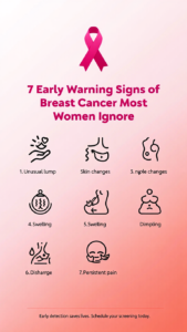
7 Early Warning Signs of Breast Cancer Most Women Dismiss
Format A: Signs You May Be Missing, with Clinical Insight for Each
Sign 1: A Change in Breast Skin Texture, Especially Puckering or Dimpling
This is one of the most clinically telling signs of early breast cancer, and it is one of the most frequently overlooked.
When a tumour grows within breast tissue, it can attach to the Cooper’s ligaments, the fibrous bands that connect the breast skin to the underlying chest wall. As the tumour grows and these ligaments are pulled inward, the skin above begins to pucker, dimple, or wrinkle in a localised area. It can look like a tiny indentation, or it can resemble the texture of an orange peel. In medical terms, the orange peel appearance is called peau d’orange, and it is often associated with inflammatory breast cancer, a particularly fast-moving type.
The reason this gets dismissed is that many women assume skin texture changes on the breast are cosmetic, related to weight fluctuation, or simply a feature of ageing skin. Sometimes they are. But if you notice a new dimple, pucker, or uneven skin surface that was not there before, and it does not resolve after your next menstrual cycle, that change warrants clinical attention. This is not about causing alarm. It is about recognising that your skin is a window into what is happening underneath it.
The key word in all of this is “new.” Longstanding asymmetry or texture that has always been present is far less concerning than something that has changed in the last few weeks or months.
Sign 2: Nipple Inversion or a Change in Nipple Direction
Nipple inversion, where the nipple turns inward or points in a different direction than it previously did, is another sign that many women rationalise away.
Some women have had inverted nipples their entire lives. That is a normal anatomical variant and generally not a cause for concern. The sign that matters here is a nipple that was previously pointing outward and has recently turned inward, or one that has changed its direction or position relative to the other. When a tumour grows behind or beneath the nipple, it can physically pull the nipple inward as the tissue retracts.
A change in nipple direction can also be accompanied by a subtle flattening of the nipple, or one nipple sitting noticeably higher than the other when viewed in a mirror. Again, the word “new” is doing all the clinical heavy lifting. If your nipples have always been slightly asymmetrical, that history matters. If this is a recent change, it belongs in the conversation with your doctor.
Do not wait and see with this one. Nipple changes are often among the earliest visible signs of breast cancer because the nipple sits at the convergence of the ductal system, where many breast cancers begin.
Sign 3: Nipple Discharge That Is Not Related to Breastfeeding
Nipple discharge outside of pregnancy or breastfeeding can feel embarrassing to mention, and many women simply do not bring it up. This is one of the communication gaps I want to close directly.
Not all nipple discharge indicates breast cancer. Milky discharge, particularly from both nipples, is more commonly related to elevated prolactin levels (a hormone that can be raised by certain medications, thyroid conditions, or a benign pituitary tumour). However, discharge that is bloody, clear, or pink, comes from only one nipple, and occurs spontaneously (without squeezing), is a symptom that requires prompt investigation.
The mechanism is important to understand. Certain breast cancers, particularly those that originate within the ducts, can bleed or produce fluid as they grow. That fluid has to go somewhere, and the path of least resistance is often through the nipple. This is why single-nipple, spontaneous, or blood-tinged discharge is taken seriously clinically, even when a lump cannot yet be felt.
A small but important nuance: clear or slightly cloudy discharge produced only when the nipple is actively squeezed is far less concerning than discharge that appears on its own in your bra or clothing. The difference matters, so be specific when you describe it to your doctor.
Sign 4: Persistent, Localised Breast Pain That Is Not Cyclical
Here is the statement that surprises many women in my clinic: most breast pain is not cancer. Cyclical breast pain tied to your menstrual cycle, tenderness that rises and falls with your hormonal rhythm, is extremely common and overwhelmingly benign.
The pain pattern that warrants investigation is different. It is one-sided. It is localised to a specific spot rather than spread across the whole breast. It does not fluctuate with your cycle. It persists for more than a few weeks. And it may be described less as tenderness and more as a dull ache, a pressure, or an intermittent burning sensation in one fixed area.
This type of non-cyclical breast pain accounts for a smaller proportion of breast pain overall, but it is one of the presentations that clinical experience tells me gets most readily dismissed, both by patients and sometimes by clinicians. When a patient tells me she has had the same aching spot in her breast for six weeks and her cycle has come and gone twice without it changing, I want to know more. Not because pain alone is a definitive sign of cancer, but because a persistent, localised symptom in breast tissue deserves a proper assessment, not reassurance based on nothing.
Track your pain. Note whether it moves, whether it changes with your cycle, and whether it stays localised to one area. That history is genuinely useful clinical information.
Sign 5: Swelling or a Feeling of Fullness in the Armpit or Around the Collarbone
The lymphatic system is the body’s biological alert network. Lymph nodes, small bean-shaped glands found in clusters throughout the body, are among the first places breast cancer spreads when it moves beyond the original tumour. The lymph nodes closest to the breast sit in the armpit (axillary nodes) and around the collarbone (supraclavicular nodes).
When breast cancer cells travel to these nodes, the nodes can enlarge, becoming palpable as a lump, or causing a feeling of fullness, tightness, or aching in the armpit, even when there is no palpable mass in the breast itself. This is a sign that frequently catches people off guard, because they are not looking for symptoms in their armpit when they think about breast cancer.
Women often mistake swollen axillary lymph nodes for a pulled muscle, an ingrown hair, or general soreness from exercise. And to be fair, those are far more common explanations. Lymph nodes also enlarge in response to infection anywhere in the upper body. The pattern that raises clinical flags is a node that is firm rather than soft, does not resolve within two to three weeks, is painless (though not always), and is not accompanied by any clear infection or injury explanation.
As I’ve seen with many patients, swelling in the armpit is often the first tangible sign a woman notices, precisely because she is not examining that area as part of a standard self-check. Start including your armpits and the area up to your collarbone in every breast self-examination.
Sign 6: A Change in Breast Size or Shape, Particularly on One Side Only
Breasts are not perfectly symmetrical. Almost no woman has two identical breasts, and lifelong mild asymmetry is entirely normal. What is worth paying attention to is a change from your personal baseline.
A breast that has recently become noticeably larger on one side, without a corresponding change in weight or hormonal status, may be exhibiting swelling related to lymphatic obstruction or tumour growth. Conversely, a breast that appears to be shrinking, sitting differently, or pulling upward in a way that does not match the other side may be showing signs of retraction related to tumour involvement of deeper tissue.
The key is comparison over time. Photographs taken regularly (once a month is sufficient) can be genuinely useful for tracking changes you might not notice day to day in the mirror. Stand in good lighting, with your arms raised and then lowered, and look for any new asymmetry in the outline, position, or volume of each breast.
One-sided breast changes that evolve over a period of four to eight weeks and are not explained by a clear hormonal shift, weight change, or injury deserve a clinical review. You do not need to be certain something is wrong to ask for an assessment. You simply need to have noticed a change.
Sign 7: Skin Redness, Warmth, or a Rash on the Breast That Does Not Resolve
Breast skin that is red, warm, thickened, or has developed a rash-like appearance is sometimes the first visible presentation of inflammatory breast cancer, one of the rarer but more aggressive forms of the disease.
Inflammatory breast cancer (IBC) is particularly challenging because it does not typically present with a discrete lump. Instead, it causes a diffuse change across the breast: redness that spreads, warmth to the touch, heaviness, rapid enlargement of the breast, and that characteristic peau d’orange texture. Because these symptoms resemble mastitis (a breast infection common in breastfeeding women) or eczema, they are sometimes treated as infections for weeks before a biopsy is considered.
The distinction matters because IBC moves quickly, and delays in diagnosis significantly affect outcomes. The clinical rule I apply is this: if breast skin redness and warmth do not respond to a course of antibiotics within one to two weeks, or if they appear in a woman who is not breastfeeding with no obvious skin condition to explain them, further investigation is not optional. It is necessary.
A rash or persistent red patch on or around the nipple area specifically should also prompt a conversation with your doctor, as it can occasionally be a sign of Paget’s disease of the nipple, a rare form of breast cancer that begins in the nipple ducts and spreads to the areola.
According to the Mayo Clinic’s complete breast cancer symptoms guide, these skin and nipple changes are among the most commonly underreported early indicators that women present with during diagnosis.
The Clinical Insight: What I Have Seen in 19 Years of Practice
In my clinical experience, the single most consistent pattern I have observed is not a failure to notice symptoms. It is a failure to act on them.
Women are remarkably attuned to their bodies. They notice things. They feel the change in texture. They register the new dimple. They mention to their partner that one breast feels different. And then, more often than I would like to report, they talk themselves out of it. They tell themselves they are being hypochondriacal. They wait until after Christmas, after the school run, after the work deadline resolves. They worry about wasting a doctor’s time.
This pattern has a name in behavioural health research. It is called appraisal delay, the gap between noticing a symptom and deciding it warrants medical attention. Studies consistently show that appraisal delay is one of the primary contributors to late-stage breast cancer diagnosis, and it is overwhelmingly driven not by a lack of knowledge about what a lump feels like, but by ambiguity about whether a symptom is serious enough to act on.
The women I see who are diagnosed at earlier, more treatable stages are not the ones who knew more about breast cancer in the abstract. They are the ones who had a clear, specific benchmark for when to act. This is what I want to give you today. Not a reason to panic. A reason to act promptly, calmly, and with the confidence that your concern is clinically valid.
Because it is. Your instinct that something has changed is worth a conversation with a doctor. Every single time.
When to See a Specialist: Specific Red Flags and Timeframes
The guidance below is not a substitute for an annual breast examination with your GP or primary care provider. It is designed to tell you when to seek attention sooner than your next scheduled appointment.
If you notice nipple discharge that is bloody, pink, or clear, coming from one nipple only, and occurring without pressure being applied: do not wait for a routine appointment. Contact your GP or gynaecologist within one week and specifically describe the discharge as spontaneous and unilateral (from one side). You may be referred for a ductogram or breast ultrasound.
If you notice skin dimpling, puckering, or a peau d’orange texture in any part of the breast: book an urgent appointment with your GP within one to two weeks. Be prepared to have a breast examination and potentially a referral to a breast surgery specialist or breast clinic.
If you have a new, localised breast pain that has persisted beyond two full menstrual cycles without changing: see your GP and ask specifically for a clinical breast examination. If you are over 35, ask whether a mammogram or ultrasound is appropriate.
If you notice a swelling or firm lump in your armpit that has been present for more than three weeks with no obvious infection or injury to explain it: request an urgent review with your GP. If a lymph node concern is confirmed on examination, a referral to a breast surgeon or general surgeon is standard.
If you have breast redness and warmth that has not improved after a seven to ten day course of antibiotics, or that is present in a woman who is not currently breastfeeding: this requires a referral to a breast specialist promptly. Specifically request that inflammatory breast cancer be excluded before further antibiotic courses are prescribed.
If you notice any new, unexplained change in breast size, shape, or nipple position that has evolved over four to eight weeks: a mammogram (for women over 40) or ultrasound (for younger women with denser breast tissue) is a reasonable and appropriate next step to request.
You do not need a dramatic symptom to deserve a clinical assessment. A change from your normal is enough.
Understanding Your Breast Density and Why It Matters for Detection
The Hidden Factor in Early Detection
This is a conversation that does not happen often enough in routine care, and it is one that directly affects how reliably early warning signs of breast cancer will show up on imaging.
Breast density refers to the proportion of glandular and fibrous tissue relative to fatty tissue in the breast. Dense breast tissue appears white on a mammogram. So does early tumour tissue. This overlap is not theoretical. Research published across multiple large breast cancer cohorts has shown that mammography is significantly less sensitive in women with dense breast tissue, with cancer detection rates dropping substantially in the highest density categories.
What this means practically is that for women with dense breasts, a normal mammogram result is genuinely reassuring but not completely definitive. Supplemental imaging, such as breast ultrasound or MRI, may be recommended for women with dense tissue who have additional risk factors. This is a conversation you can and should initiate with your radiologist or GP if you have had a mammogram and were told you have dense breast tissue.
Knowing your breast density is not a reason for anxiety. It is a reason to have a more nuanced conversation about your screening protocol and to be especially attentive to the physical and visual changes described throughout this article.
How to Perform an Effective Breast Self-Examination
A breast self-examination (BSE) is not a diagnostic test. It will not replace imaging. But performed consistently and correctly, it is an invaluable tool for establishing your personal baseline, the specific topography of your own breasts, so that changes become detectable.
Here is the correct technique:
In the shower: Using the pads (not the tips) of your three middle fingers, move in small circular motions across the entire breast, including the armpit and the area below the collarbone. Use three levels of pressure: light, medium, and firm. Cover the full breast in a grid pattern from top to bottom, or in a circular motion from the outside in toward the nipple.
In the mirror: Stand with your arms at your sides, then raise them overhead. Look for any new asymmetry in size, shape, skin texture, nipple position, or the outline of each breast. Then place your hands on your hips and gently press inward to flex the chest muscles, and observe again.
Lying down: Place a pillow under your right shoulder and your right arm behind your head. Use your left hand to examine the right breast using the same circular, three-pressure technique. Repeat on the other side.
The best time to perform a breast self-examination is seven to ten days after the start of your period, when hormonal breast swelling and tenderness are at their lowest. If you are post-menopausal or not menstruating, choose one consistent day of the month and examine on that date every month.
Who Is at Higher Risk and What You Can Do About It
Risk Factors That Deserve Your Attention
Understanding your personal risk profile for breast cancer does not mean living in fear. It means making informed decisions about screening frequency, lifestyle, and whether genetic counselling is appropriate for you.
Several well-established risk factors increase a woman’s likelihood of developing breast cancer across her lifetime.
Family history is one of the most significant. Having a first-degree relative (mother, sister, or daughter) who has been diagnosed with breast cancer raises your lifetime risk. Having multiple relatives affected, particularly those diagnosed before the age of 50, or having a relative with ovarian cancer, raises the possibility of an inherited gene mutation (most commonly BRCA1 or BRCA2) and may warrant genetic testing and counselling.
Age remains a primary risk factor. The majority of breast cancers are diagnosed in women over the age of 50. However, as I see in my clinic with increasing frequency, breast cancer in younger women (under 40) does occur, and it is often diagnosed at a later stage because younger women are less likely to be screened and more likely to attribute symptoms to hormonal causes.
Dense breast tissue, as discussed above, both increases risk slightly and reduces mammographic sensitivity.
Hormonal factors including early onset of menstruation (before age 12), late menopause (after age 55), not having had children, or having a first pregnancy after the age of 30 are all associated with a modest increase in breast cancer risk. These reflect cumulative lifetime exposure to oestrogen, which can promote the growth of certain breast cancer cell types.
Lifestyle factors including alcohol consumption, being overweight after menopause (because fatty tissue produces oestrogen), and physical inactivity are modifiable risk factors. According to research from the National Institutes of Health (NIH) on breast cancer risk factors, even moderate alcohol intake (one drink per day) is associated with a small but measurable increase in breast cancer risk, which is worth factoring into informed personal decisions.
None of these factors are a guarantee. Women with no identifiable risk factors develop breast cancer. Women with multiple risk factors do not. Risk is not destiny. But it is useful information.
The Importance of Regular Screening
Current clinical consensus recommends that women with average risk begin annual or biennial mammographic screening from the age of 40 to 50, depending on national guidelines and individual factors. Women with elevated risk due to family history or genetic mutations may be recommended to begin screening earlier, potentially from the age of 25 to 30, and may be offered MRI in addition to mammography.
If you are unsure what screening schedule is right for you, that conversation belongs in your next GP or gynaecology appointment. Ask directly: based on my age, family history, and breast density, what screening do you recommend, and how often?
That question takes thirty seconds to ask. The answer could change everything.
Separating Fear From Fact: Common Myths About Breast Cancer Signs
Myth 1: If There Is No Lump, There Is No Cancer
This is arguably the most harmful myth in breast cancer awareness. As this article has detailed, several forms of breast cancer, including inflammatory breast cancer and some lobular carcinomas, may present without a palpable lump for a significant period. Nipple changes, skin texture alterations, lymph node swelling, and breast asymmetry can all be early signs of breast cancer in the complete absence of any detectable mass.
The goal of a breast self-examination is not just to find lumps. It is to notice anything that is different from your established baseline.
Myth 2: Breast Pain Usually Means Cancer
The reverse of the above is equally important to correct. Most breast pain is not cancer. Cyclical mastalgia (pain linked to the menstrual cycle) is extremely common and almost always benign. Non-cyclical breast pain also has many benign causes, including fibrocystic breast changes, costochondritis (inflammation of the cartilage connecting the ribs to the breastbone), and musculoskeletal strain.
Pain alone, particularly when it is bilateral (in both breasts) and fluctuates with the menstrual cycle, is a low-risk symptom. It is the persistence, localisation, and one-sided nature of breast pain that raises the clinical index of suspicion.
Myth 3: Only Older Women Need to Check Regularly
Breast cancer does occur in younger women, and as I mentioned earlier, it is frequently diagnosed at a later stage in younger age groups precisely because screening programmes do not typically begin until age 40 to 50 and because younger women are less likely to present promptly with symptoms.
Women in their 20s and 30s benefit from monthly breast self-examination and should report any persistent changes to their GP regardless of age. Knowing your baseline now makes detecting a change in the future far more reliable.
Myth 4: A Mammogram Would Have Found It Already
Mammography is an excellent screening tool, but it is not perfect, particularly in women with dense breast tissue. False negatives do occur. This is not a criticism of mammography, it remains the gold standard for population screening, but it underscores why clinical breast awareness and self-examination remain valuable complements to imaging.
If you notice a change between scheduled mammograms, do not wait until your next one. Present promptly and request a clinical assessment.
Myth 5: Breast Cancer Always Runs in Families
Approximately 85 to 90 per cent of breast cancer cases occur in women with no family history of the disease. The majority of breast cancers are sporadic, meaning they are not driven by an inherited mutation but by a complex interaction of hormonal, environmental, and cellular factors over time.
Family history matters and should be shared with your healthcare provider. But the absence of family history is not a reason to become complacent about breast awareness. Breast cancer is the most common cancer diagnosed in women worldwide, and most women who develop it have no close relatives with the disease.
A Note on Emotional Weight: The Psychology of Breast Symptoms
There is something I want to name directly, because it matters enormously and rarely gets clinical airtime.
The fear of breast cancer is real. It is culturally embedded, socially reinforced, and for many women, emotionally laden with personal grief, whether from a mother’s diagnosis, a friend’s treatment, or their own previous scare. That fear is valid.
And paradoxically, it can work in both directions. It can motivate prompt action, or it can trigger avoidance. Some women present at the first possible hint of a symptom. Others delay for months because they cannot face the possibility of what a doctor might find.
If you recognise yourself in the second description, I want to be clear with you: avoiding the appointment does not make the symptom go away. It simply reduces the window for early intervention. And early intervention, when it comes to breast cancer, is where outcomes are genuinely transformed.
The anxiety of not knowing is almost always harder to carry than the clarity of a definitive answer. Most breast symptoms, even when investigated, turn out to have benign explanations. You will walk out of that appointment knowing more than you walked in. That is always a better position to be in.
Go. Make the appointment. You deserve clarity.
Building a Proactive Breast Health Routine
A Monthly Practice That Could Save Your Life
Building a consistent breast awareness routine is one of the most practical and empowering things you can do for your long-term health. Here is a realistic monthly framework:
Day 7 to 10 after your period starts: Perform a thorough breast self-examination using the three-position technique described earlier (in the shower, in the mirror, lying down). If you are post-menopausal, choose a fixed calendar date each month.
Monthly: Take a photograph in good lighting with arms raised and arms lowered. Store these privately. Review them over time rather than immediately, as subtle changes are easier to identify when comparing images from three to six months apart rather than week to week.
Annually: Attend your scheduled mammogram if you are in the screened age group, and have a clinical breast examination performed by your GP or gynaecologist. Bring notes. Mention any changes you have noticed, no matter how minor they seem.
Whenever you notice a change: Do not apply a wait-and-see approach beyond one full menstrual cycle for most symptoms, and immediately for blood-tinged nipple discharge or rapidly developing breast skin changes.
Consistency is what creates clinical value here. A self-examination performed once a year is of limited use. Performed monthly, it builds the intimate knowledge of your own anatomy that makes early detection possible.
What Happens If You Are Referred to a Breast Clinic
Many women feel profound anxiety at the words “I am going to refer you to the breast clinic.” It is worth clarifying what that referral typically means.
A referral to a breast clinic, particularly an urgent referral, does not mean your doctor believes you have cancer. In many health systems, breast clinic referrals are triggered by any symptom that warrants specialist assessment. The vast majority of women referred to breast clinics do not receive a cancer diagnosis.
At a breast clinic appointment, you will typically receive a clinical breast examination by a specialist, followed by imaging (mammogram, ultrasound, or both, depending on your age and symptoms), and occasionally a needle biopsy if there is an area of tissue that needs closer examination. This is called triple assessment: clinical examination, imaging, and pathology if required.
The process is designed to be thorough and reassuring, not alarming. Most appointments result in a clear explanation of what the symptom was (often a benign cyst, fibrocystic change, or normal hormonal tissue variation) and a return to routine screening.
Knowing this in advance can reduce the emotional weight of the referral significantly. It is a thorough clinical process, not a verdict.
Empowering You to Act: Your Next Step Starts Today
You have just done something important. You have spent time actively learning about your body, about the specific signals it might send, and about the difference between what warrants attention and what does not. That knowledge has genuine clinical value.
The single most important takeaway from everything in this article is this: a change from your personal baseline, in any of the seven areas we have covered, is a reason to make an appointment. Not a reason to panic. A reason to act with calm, informed confidence.
You do not need to have a perfect self-examination technique before you start. You do not need a dramatic symptom to deserve medical attention. You need to know your body well enough to notice when something has shifted.
Start this month. Set a reminder in your phone for day eight after your next period. Take five minutes. Learn what is normal for you. And if something feels or looks different, say so out loud to your doctor. Use the specific language from this article if it helps: “I have noticed a new dimple,” “I have persistent localised pain in one area that is not related to my cycle,” “There is a firmness in my armpit that has been there for three weeks.”
That specificity is what moves clinical consultations forward. That is how early detection happens.
Share this article with a woman in your life who needs clear, trustworthy information about her breast health. It may be the most useful thing she reads this year.
Medical Disclaimer
This article is for informational purposes only and does not constitute medical advice. Always consult a qualified healthcare provider before making any changes to your health or treatment plan.
Dr. Naomi is a board-certified women’s health physician with 19 years of clinical experience in reproductive endocrinology and integrative gynaecology. She writes exclusively for webzalo.com, a trusted women’s health resource for women aged 25 to 50 navigating reproductive and hormonal health challenges.

